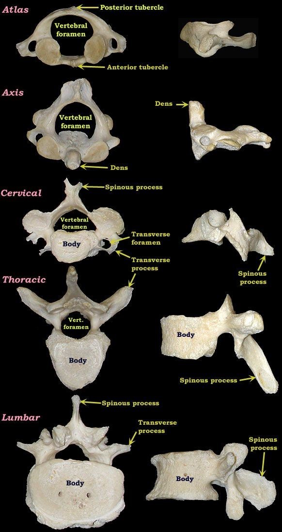
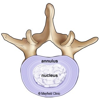
• Discs are designed like a radial car tire. The outer ring, called the annulus, has crisscrossing fibrous bands, much like a tire tread.
• These bands attach between the bodies of each vertebra. Inside the disc is a gel-filled centre called the nucleus, much like a tire tube
• Discs function like coiled springs. The crisscrossing fibers of the annulus pull the vertebral bones together against the elastic resistance of the gel-filled nucleus.
• The nucleus acts like a ball bearing when you move, allowing the vertebral bodies to roll over the incompressible gel. The gel-filled nucleus contains mostly fluid.
• This fluid is absorbed during the night as you lie down and is pushed out during the day as you move upright.
• With age, our discs increasingly lose the ability to reabsorb fluid and become brittle and flatter; this is why we get shorter as we grow older.
• Also diseases, such as osteoarthritis and osteoporosis, cause bone spurs (osteophytes) to grow.
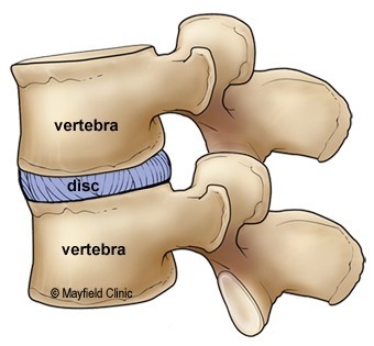
• Injury and strain can cause discs to bulge or herniate, a condition in which the nucleus is pushed out through the annulus to compress the nerve roots causing back pain.
Muscles
• The two main muscle groups that affect the spine are extensors and flexors.
• The extensor muscles enable us to stand up and lift objects. The extensors are attached to the back of the spine.
• The flexor muscles are in the front and include the abdominal muscles.
• These muscles enable us to flex, or bend forward, and are important in lifting and controlling the arch in the lower back.
• The back muscles stabilize your spine. Something as common as poor muscle tone or a large belly can pull your entire body out of alignment. Misalignment puts incredible strain on the spine.
Facet joints
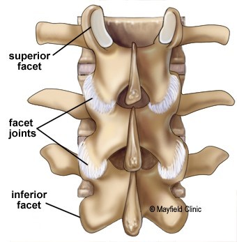
The facet joints of the spine allow back motion. Each vertebra has four facet joints, one pair that connects to the vertebra above (superior facets) and one pair that connects to the vertebra below (inferior facets).
Spinal cord
• The spinal cord is about 18 inches long and is the thickness of your thumb.
• It runs from the brainstem to the 1st lumbar vertebra protected within the spinal canal.
• At the end of the spinal cord, the cord fibers separate into the cauda equina and continue down through the spinal canal to your tailbone before branching off to your legs and feet.
• The spinal cord serves as an information super-highway, relaying messages between the brain and the body.
• The brain sends motor messages to the limbs and body through the spinal cord allowing for movement. The limbs and body send sensory messages to the brain through the spinal cord about what we feel and touch. Sometimes the spinal cord can react without sending information to the brain.
• These special pathways, called spinal reflexes, are designed to immediately protect our body from harm.
• Any damage to the spinal cord can result in a loss of sensory and motor function below the level of injury.
• For example, an injury to the thoracic or lumbar area may cause motor and sensory loss of the legs and trunk (called paraplegia). An injury to the cervical (neck) area may cause sensory and motor loss of the arms and legs (called tetraplegia, formerly known as quadriplegia).
Spinal nerves
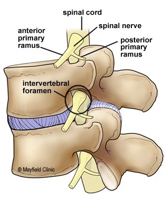
• Thirty-one pairs of spinal nerves branch off the spinal cord. The spinal nerves act as “telephone lines,” carrying messages back and forth between your body and spinal cord to control sensation and movement.
• Each spinal nerve has two roots. The ventral (front) root carries motor impulses from the brain and the dorsal (back) root carries sensory impulses to the brain.
• The ventral and dorsal roots fuse together to form a spinal nerve, which travels down the spinal canal, alongside the cord, until it reaches its exit hole - the intervertebral foramen.
• Once the nerve passes through the intervertebral foramen, it branches; each branch has both motor and sensory fibers.
• The smaller branch (called the posterior primary ramus) turns posteriorly to supply the skin and muscles of the back of the body. The larger branch (called the anterior primary ramus) turns anteriorly to supply the skin and muscles of the front of the body and forms most of the major nerves.